

Proactive Mall MRI screenings

Mall MRI Screening Services: The New Era of Proactive Health in the USA
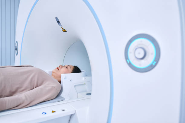
The Rise of Mall and Retail MRI Screening
In recent years, the United States has experienced the rapid growth of retail and mall-based MRI screening services. Companies such as Prenuvo and Ezra now offer whole-body MRI scans at several urban centers and upscale malls, providing consumers with unprecedented access to this advanced diagnostic technology. These facilities attract people who desire proactive control of their health, opting to pay out of pocket rather than navigate insurance barriers. The trend is driven by frustrations over insurance companies refusing to cover MRI’s without a specialist referral, and doctors often discouraging “unnecessary” screenings if patients are symptom-free. As a result, empowered individuals are turning to these services for early detection—especially for peace of mind regarding cancer risk.ezra+1
Cancers That MRI’s Can Detect
MRI scans are most effective at detecting cancers in soft tissues and organs. The following is a numbered list of cancers that MRI’s are equipped to detect, often at early stages:
- Brain cancerglmi+1
- Spinal cancerhealthline
- Breast cancer (especially effective for high-risk individuals and those with dense breast tissue)glmi+1
- Prostate cancerhealthline+1
- Liver cancer (hepatocellular carcinoma)glmi+1
- Pancreatic cancerhealthline+1
- Ovarian cancerglmi+1
- Bladder cancerhealthline
- Lung cancer (especially for assessing tumor spread and location)healthline
- Esophageal cancerhealthline
- Multiple myelomahealthline
- Non-Hodgkin’s lymphomahealthline
These cancers are listed due to MRI’s superior ability to visualize abnormal growths, soft tissue masses, and lesions in these areas.
Cancers Less Likely to Be Detected by MRI
While MRI is a powerful imaging tool, there are certain cancers and circumstances where its effectiveness is limited:
- Small or early-stage tumors in organs with motion (e.g., colon, stomach)healthline
- Blood cancers (e.g., leukemia), which are systemic and do not form solid masses
- Some forms of skin cancer (e.g., basal and squamous cell carcinoma)
- Cancers confined to mucosal surfaces or very early lesions in the GI tract
- Certain low-grade prostate cancers, especially small or non-aggressive types, can sometimes be missed by MRI (false negatives)pmc.ncbi.nlm.nih+1
- Kidney and testicular cancers, where ultrasound or CT may be better for small lesions
It’s important to note that while MRI is valuable for detecting tumors, not all cancers form discrete masses visible on MRI, and no screening tool is perfect.
MRI: History, Invention & Key Dates
- The principle behind MRI technology (nuclear magnetic resonance) was discovered in the 1940s by Felix Bloch and Edward Purcell.
- The actual mechanism to create imaging from NMR was developed by Paul C. Lauterbur in September 1971, who published on spatial encoding with magnetic field gradients in 1973.wikipedia+2
- Peter Mansfield further refined image acquisition and processing techniques. Both Lauterbur and Mansfield were awarded the Nobel Prize in Physiology or Medicine in 2003 for their contributions to MRI.stonybrook+1
- Dr. Raymond Damadian filed the first MRI patent in 1972, built one of the first NMR scanning machines (“Indomitable”), and performed the first full-body human MRI scan in 1977.ezra+1
Types of MRI Scans & Their Differences
MRI technology includes a variety of scan types used for different diagnostic purposes:
- Standard/Anatomic MRI: Produces detailed anatomical images using T1-weighted and T2-weighted sequences.
- Functional MRI (fMRI): Measures brain activity by detecting changes in blood flow, used in neuroscience research and surgical planning.ezra+1
- Magnetic Resonance Angiography (MRA): Specializes in detailed images of blood vessels and arteries, used to detect vascular conditions.innovativemri+1
- Diffusion MRI (dMRI): Maps the diffusion of water molecules in tissues, most valuable in acute stroke diagnosis and white matter brain imaging.ezra
- Cardiac MRI: Focuses on heart structures and function, identifies heart muscle damage and vascular abnormalities.innovativemri
- Breast MRI: Used for high-risk breast cancer screening and for clarifying uncertain findings from mammography or ultrasound.innovativemri
- Bone and Joint MRI: Visualizes muscles, ligaments, tendons, cartilage, and bone marrow, ideal for sports injuries and bone tumors.brownhealth+1
- Abdominal and Pelvic MRI: Examines specific organs like the liver, kidneys, uterus, or prostate for masses or abnormalities.brownhealth+1
- MR Spectroscopy: Assesses chemical composition of tissues, sometimes used to distinguish tumor types.
- Open MRI: Uses a more open design for patients with claustrophobia, though may have lower resolution compared to closed MRI systems.innovativemri
Sequence Variants
- T1-weighted: Great for anatomy.
- T2-weighted: Excellent for highlighting pathology/injury due to water content.
- Contrast-enhanced scans: Use gadolinium agents to improve lesion/tumor detection in specific clinical settings.innovativemri
Each MRI type and sequence is selected based on the organ system, clinical question, and patient tolerance.